Breast cancer remains a significant health challenge globally, and the methods used for its diagnosis are crucial in ensuring effective treatment. While mammograms are the standard screening tool for breast cancer, the role of other imaging techniques, particularly chest computed tomography (CT) scans, warrants a closer examination. Although not routinely employed for breast cancer diagnosis, CT scans can occasionally reveal incidental findings that may indicate the presence of breast cancer.
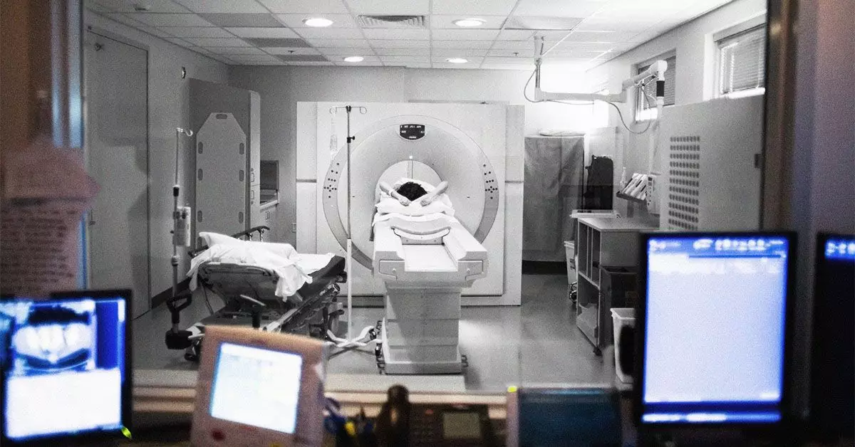
CT scans provide detailed, cross-sectional images of the body by utilizing X-rays. This imaging technique compiles multiple 2D X-ray images to form comprehensive 3D visuals of the internal structures within the body. The scan’s capability to discern various tissues, organs, and bones facilitates the identification of abnormalities. To enhance image clarity, particularly of soft tissues and blood vessels, contrast dye—commonly iodine or barium—is often introduced intravenously or orally. Such contrasts are particularly advantageous in unveiling subtle details that might otherwise go unnoticed.
Despite their detailed imaging capabilities, CT scans are not standard protocol for breast cancer screening. Research indicates that only a small percentage of cases rely solely on CT findings for breast cancer diagnosis. A notable 2021 study highlighted that while incidental findings on CT scans might suggest breast cancer, the scans themselves are not optimized for breast health assessment.
One alarming aspect of utilizing CT scans for unrelated health concerns is the potential for incidental findings, which—according to research—can indicate breast cancer in up to 28% of cases. This statistic emphasizes the importance of careful interpretation of CT visuals by healthcare professionals. Interestingly, non-contrast CT scans may outperform mammography under specific conditions, such as when lesions reside deep within dense breast tissues adjacent to the chest wall. This highlights potential overlaps in imaging efficacy that may go underappreciated in standard practice.
Nevertheless, incidental findings pose a double-edged sword. While they can lead to early detection of breast cancer, they may also subject patients to unnecessary anxiety and further investigations. The critical responsibility rests on doctors to strike a balance between vigilance and avoid overdiagnosis.
In assessing breast disease, several imaging modalities are utilized, with mammograms leading the forefront. Standard screenings employ low-dose X-rays aimed at detecting abnormalities in breast tissue with minimal radiation exposure. When abnormal findings emerge on screening mammograms, follow-up diagnostic mammograms offer more detailed imaging.
In addition to mammograms, healthcare providers often employ complementary tests like breast ultrasounds, MRI scans, and biopsies. Ultrasound relies on sound waves to create images, while MRI utilizes magnets and radio waves, offering greater detail in soft tissue visualization. A biopsy, on the other hand, provides definitive diagnosis by extracting tissue samples for pathological examination.
Unlike these focused breast cancer diagnostic tools, CT scans are primarily used to evaluate conditions across various body systems. They play a pivotal role in determining whether breast cancer has metastasized to other organs, such as the lungs and liver. This broader context highlights the significance of CT imaging as part of an integrated diagnostic strategy rather than a standalone approach to breast cancer diagnosis.
The possibility that a routine chest CT scan could uncover incidental findings of breast cancer reinforces the need for continuous healthcare provider education. It is imperative that medical professionals review CT images with a discerning eye for breast anomalies, given that these discrepancies could significantly impact patient outcomes. A 2023 study indicated that radiologists miss about 64.3% of incidental breast cancer findings on chest CT scans, demonstrating a critical gap in diagnostic accuracy that must be addressed.
Moreover, patients themselves must be vigilant. Any new alterations, such as lumps or changes in breast tissue or the nipple, should prompt discussions with healthcare providers. Early intervention remains paramount in enhancing survival rates, and patients should be encouraged to participate actively in their health monitoring.
While chest CT scans are not traditional diagnostic tools for breast cancer screening, their potential for revealing incidental findings cannot be overlooked. As healthcare practices evolve, the integration of various imaging modalities, including CT scans, must be considered within a broader strategy for early detection and treatment. Patients and healthcare providers alike need to remain proactive and informed, ensuring that breast cancer is diagnosed as early as possible, thus improving treatment outcomes across the board.

