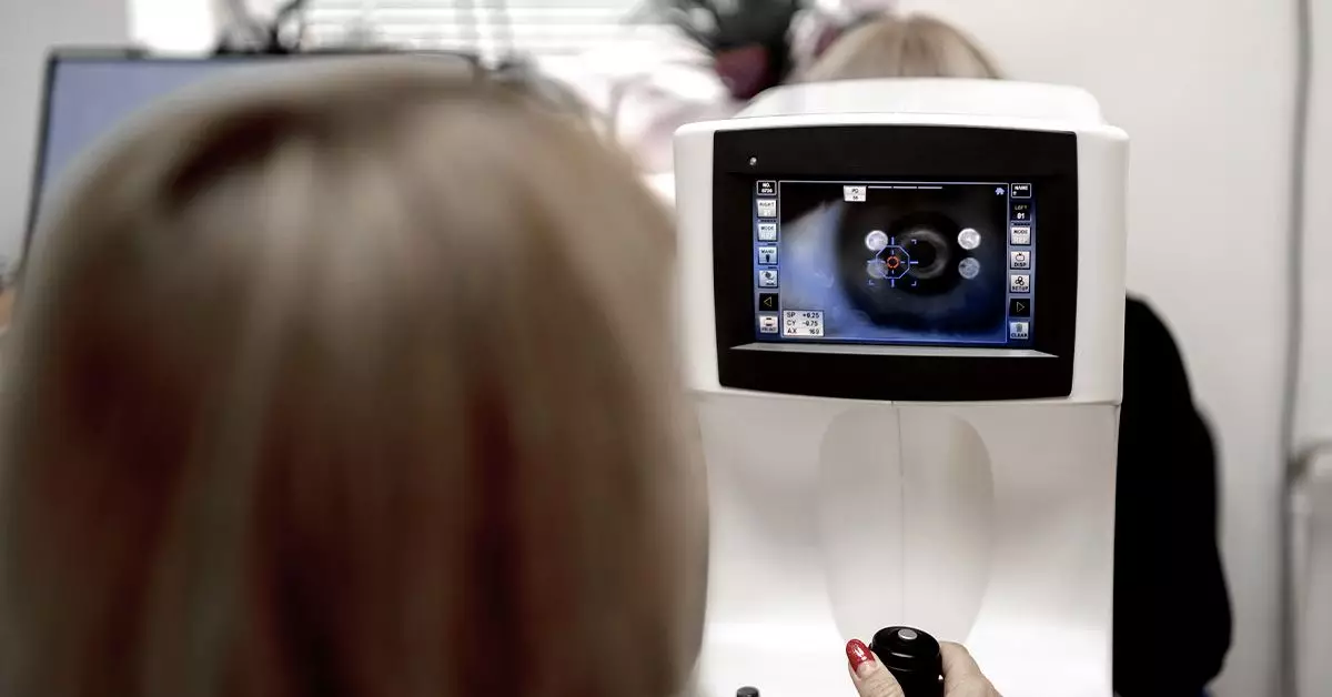
Fundus photography stands out as a vital diagnostic technique employed in the realm of ophthalmology, particularly for those grappling with diabetes. This specialized imaging process captures detailed photographs of the retina, the inner layer of the eye, allowing healthcare providers to identify various retinal conditions, notably diabetic retinopathy. The significance of this technique cannot be understated, as diabetic retinopathy remains one of the leading causes of blindness among adults worldwide. By leveraging the advantages of fundus photography, early detection and management of this condition become increasingly possible, safeguarding patients against the long-term consequences of sight impairment.
Understanding diabetic retinopathy as a progressive eye disease is crucial for both patients and healthcare providers. It arises as a complication of diabetes, particularly for those who struggle to maintain optimal blood sugar levels. Initially, the condition often develops silently, without any noticeable symptoms. However, as the disease advances, it can cause significant visual disturbances and ultimately lead to blindness if not accurately diagnosed and treated. Regular screenings through fundus photography can be instrumental in catching the earliest signs of this condition, potentially averting irreversible damage to one’s vision.
The process by which the disease manifests involves changes to the retinal blood vessels. These alterations include the formation of microaneurysms, the proliferation of new blood vessels, and the development of retinal hemorrhages. Fundus photography is designed to reveal these specific anomalies, such as microaneurysms appearing as tiny red dots or hemorrhages manifesting as dark patches within the retina. Recognizing these early indicators can facilitate timely interventions, ranging from monitoring to more aggressive treatments, such as laser therapy or anti-VEGF injections.
Recent advancements in technology have broadened the horizon of how we can conduct fundus photography. Selfie fundus imaging (SFI) represents a groundbreaking development, empowering individuals to take retinal photos using their smartphones. This innovative approach enables patients to capture images of their retinas with relative ease, only requiring the application of dilating eye drops beforehand. A study conducted in 2022 assessed the accuracy and reliability of SFI compared to traditional methods, revealing promising results. Participants who took their own smartphone photos achieved results comparable to those taken by trained technicians, thereby increasing accessibility to critical eye examinations for many individuals with diabetes.
This shift toward SFI not only streamlines the process of monitoring eye health, but also significantly reduces costs associated with traditional methods. With less reliance on specialized equipment and the ability for patients to engage directly in their healthcare, SFI has the potential to democratize access to critical screenings, especially in underserved communities where traditional ophthalmologic services may be in short supply.
While the procedure of fundus photography itself is relatively straightforward and painless, it does require some preparatory steps. Patients are commonly advised to use topical solutions for pupil dilation, a process that can lead to temporarily blurred vision and increased sensitivity to light. After undergoing the procedure, individuals are cautioned against driving until their vision returns to normal, which usually happens within a few hours. The overall screening process is efficient, with the photography taking only a few minutes, but patients must account for the time needed for the eye drops to take effect.
For those managing diabetes, the recommendation is clear: regular dilated eye exams should be performed at least annually. This frequency allows medical professionals to track any changes over time and intervene early if necessary. Particularly critical is the timing for pregnant women with diabetes, who should schedule an eye exam soon after becoming aware of their condition to ensure that any potential complications are addressed promptly.
For many patients, the primary challenge lies in the asymptomatic nature of early-stage diabetic retinopathy. However, as the condition progresses, symptoms can include fluctuations in vision, the appearance of floaters, or occasional difficulty focusing on distant objects. Such developments signal a need for immediate consultation with a healthcare professional. By maintaining vigilance and being proactive about eye health, individuals with diabetes can navigate this potential threat to their vision more effectively.
Fundus photography serves as an indispensable tool in the combat against diabetic retinopathy. By facilitating early detection and continuous monitoring, this imaging technique plays a pivotal role in preserving vision for those living with diabetes. Coupled with innovative technologies like SFI, the future of diabetic eye care looks promising, with the potential for improved accessibility and better patient outcomes. To mitigate the risk of blindness, individuals are encouraged to prioritize regular eye examinations and engage in proactive management strategies, such as maintaining glycemic control through diet, exercise, and medication. Regular screenings paired with responsible health management can together ensure a clearer path toward preserving sight for the future.

